Fetal Anomaly Scanning: What Expectant Parents Should Know
Dr. Shane Khan · 9 June 2026

For many expectant parents, the fetal anomaly scan is one of the most anticipated milestones of pregnancy. Carried out typically between 18 and 22 weeks of gestation, this detailed ultrasound examination allows us to carefully assess the developing baby’s anatomy — the brain, spine, heart, kidneys, abdominal wall, limbs, and facial structures. It is an extraordinary window into early life, and for the vast majority of families, it brings reassurance and joy.
But for a smaller number, the scan may identify findings that require further evaluation. Understanding what the scan can and cannot tell us — and how findings are communicated — is something every family deserves to know before they walk through the door.
What the Scan Involves
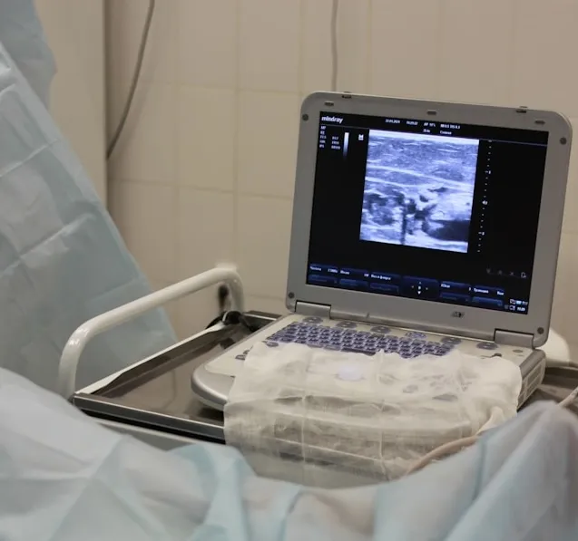
The anomaly scan is performed using high-resolution ultrasound. You will lie comfortably while the sonographer or physician passes a transducer over your abdomen, using sound waves to build a real-time image of the baby. The examination typically takes between 30 and 45 minutes, though this can vary if the baby is in an awkward position or if additional views are needed.
We examine a systematic checklist of anatomical structures. The four-chamber view of the heart, the integrity of the abdominal wall, the position of the kidneys, the shape of the head, the length and structure of all four limbs, the face — each is assessed methodically and documented.
“The anomaly scan is not a guarantee. It is a careful, skilled appraisal of the developing anatomy at a single point in time. Some findings only become apparent later in pregnancy, and some conditions are beyond the resolution of even the most detailed ultrasound.”
How Findings Are Communicated
If the scan identifies something that warrants further attention — whether a soft marker, a structural variant, or a more significant concern — you will be told clearly and compassionately, ideally by a clinician with subspecialty experience in fetal medicine. This is not a moment to rush.
In a fetal medicine setting, the conversation after an abnormal scan is as important as the scan itself. We explain what was seen, what it may or may not mean, and what the next steps are. That might involve a repeat scan in two to four weeks, a referral for fetal echocardiography, genetic counselling, or discussion of invasive testing such as amniocentesis. Every family receives individualised guidance based on their specific findings and circumstances.
It is normal to feel anxious during and after the scan, particularly if there is any uncertainty. Please know that you are not alone in that experience, and that we are here to support you.
Preparing for the Appointment
There is no special preparation required for the standard anomaly scan — you do not need a full bladder at this stage of pregnancy. Wearing comfortable, loose-fitting clothing is helpful. You are welcome to bring a partner or support person, and We encourage you to write down any questions you have beforehand, no matter how small they may seem.
If you have a history of a previous pregnancy affected by a structural anomaly, a chromosomal condition, or another complication, please let your care team know before the scan. This context helps us tailor the examination and ensures that any areas of particular concern receive the most careful attention.
A Note on Limitations
No ultrasound scan — however thorough — can detect every possible condition. The overall detection rate for major structural anomalies at the 20-week scan is high, but it is not 100%. Some conditions are simply not visible at this stage, and others lie at the boundary of what ultrasound can resolve. This is not a failure of care; it is the honest reality of the technology, and one that should be understood clearly before any scan takes place.
What we can promise is a thorough, unhurried examination performed with care — and an honest, supportive conversation about whatever we find.